Understanding Right Ventricle Dysfunction: Causes and Care


Intro
Right ventricular dysfunction (RVD) doesn’t always get the spotlight it deserves. Often overshadowed by left-sided heart failures, this condition can be a silent killer, creeping up on individuals with a series of overlooked symptoms. The right ventricle is tasked with pumping deoxygenated blood to the lungs; when it stumbles, the entire cardiovascular system feels the strain. Understanding RVD is crucial, not just for clinicians but for anyone interested in heart health.
RVD can stem from various causes—everything from pulmonary hypertension to congenital heart defects can throw a wrench in its function. In this article, we aim to put RVD under the microscope, dissecting its mechanisms and the repercussions it has on overall health. Whether you are a student scribbling notes on cardiac pathophysiology or a seasoned professional brushing up on current management strategies, the insights here will be invaluable.
Methodology
Overview of research methods used
To delve into RVD effectively, a blend of research methodologies was employed. We gathered qualitative data through systematic reviews of existing literature and quantitative data through clinical studies. This multifaceted approach ensures a comprehensive perspective, highlighting not just isolated findings but also broader trends in RVD research.
- Clinical Trials: Many recent investigations analyze treatment efficacy through randomized control trials, establishing baseline metrics for management outcomes.
- Case Studies: Detailed accounts of individual patient experiences reveal the variability in manifestation and response to treatment.
- Registry Data: National and international registries provide critical data on disease prevalence and patient demographics.
Data collection techniques
Careful data collection is a backbone of understanding any medical condition. For RVD, various techniques were applied:
- Imaging Procedures: Techniques such as echocardiograms and MRI help in visualizing the structural and functional aspects of the right ventricle.
- Biomarker Analysis: Blood tests for specific markers express inflammation and stress related to cardiac function, aiding in diagnosis.
- Patient Surveys: Gathering self-reported data from patients helps bridge the gap between clinical findings and real-world implications, reinforcing the necessity for patient-centered approaches.
Future Directions
As we march towards more advanced cardiovascular care, the future of RVD looks promising yet complex. Studies are increasingly focusing on:
Upcoming trends in research
With technology on our side, the landscape of RVD treatment is undergoing transformative changes. Research in the following areas appears particularly promising:
- Personalized Medicine: Genetic studies are paving the way for tailored treatment approaches that consider individual patient profiles.
- Wearable Technology: Devices that monitor heart function in real time could change how we track RVD progression.
- Artificial Intelligence: AI is set to revolutionize diagnostics, helping clinicians identify RVD early and accurately.
Areas requiring further investigation
Even with the strides being made, several gaps remain:
- Long-term Effects: More studies are needed to understand how RVD evolves over time, especially in younger patients.
- Integrative Approaches: Exploring how RVD impacts comorbidities is crucial for developing holistic treatment plans.
- Psychosocial Factors: Looking into the mental and emotional impacts of dealing with RVD could inform better patient support initiatives.
As we peel back the layers on right ventricular dysfunction, it's clear this issue warrants our attention now more than ever.
Understanding Right Ventricle Dysfunction
The concept of right ventricle dysfunction is not just a niche topic for specialists; it's pivotal in the broader field of cardiovascular health. Understanding this dysfunction can illuminate a variety of clinical scenarios, bridging the gap between observed symptoms and underlying pathophysiology. When we talk about right ventricle dysfunction, we are essentially looking at how this part of the heart can fail to perform its duties effectively, affecting overall heart function and leading to systemic consequences.
A comprehensive grasp of right ventricle dysfunction paves the way for early identification and management strategies that can drastically improve patient outcomes. Physicians, researchers, and educators must be well-acquainted with the nuances of this dysfunction, as it often intersects with other cardiovascular issues. Knowing how to recognize the signs, from clinical presentation to diagnostic imaging, can mean the difference between effective treatment and potential morbidity.
Definition and Overview
Right ventricle dysfunction is characterized by a decline in the ability of the right ventricle to pump blood efficiently. This may result from a host of conditions, including but not limited to structural heart abnormalities, pulmonary hypertension, or ischemic diseases. The right ventricle has a distinct role in pulmonary circulation; it transports deoxygenated blood to the lungs for oxygenation. So when it falters, there are widespread implications not only for the heart itself but also for the lungs and systemic circulation.
Essentially, when this chamber is hindered in its function, it can trigger a series of detrimental effects that lead to complications such as heart failure or arrhythmias, prompting the need for timely intervention.
Anatomical and Functional Characteristics
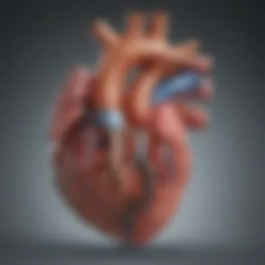
Anatomically, the right ventricle is unique in its structure compared to the left. It is thinner walled and crescent-shaped, which reflects its specific job of pushing blood into the relatively low-pressure system of the pulmonary arteries. Understanding this anatomy is crucial because it influences how diseases affect the heart's performance.
Functionally, the right ventricle must maintain a delicate balance of pressures; high pressures can lead to right ventricular hypertrophy, while low pressures can be a sign of dysfunction.
Moreover, its interaction with the left ventricle and conductance system is vital to ensure that blood flow remains uninterrupted. Factors such as volume overload, pressure overload, and myocardial ischemia can all contribute to dysfunction in this chamber. Therefore, both anatomical and functional characteristics of the right ventricle are intertwined, creating a complex network that, when disturbed, poses serious health risks.
"Understanding the intricacies of right ventricle dysfunction offers invaluable insights into the overarching mechanisms at play within the cardiovascular system."
Grasping these characteristics not only enhances clinical reasoning but allows professionals to tailor interventions that might avert further complications.
Pathophysiology of Right Ventricle Dysfunction


Understanding the pathophysiology of right ventricle dysfunction (RVD) is crucial because it lays the groundwork for recognizing its implications, not just for cardiovascular health, but also for the overall well-being of patients. It's like peeling an onion; until you get to the core, you don’t really understand the tears caused by dysfunction. The right ventricle plays a pivotal role in pumping deoxygenated blood to the lungs, and any disruption in this function can lead to cascading effects that affect various body systems.
RVD often arises from a myriad of mechanisms that change the normal functioning of the heart. It is not merely an isolated issue but rather interlinked with other cardiovascular diseases, making it vital to understand its pathophysiological underpinnings. By diving into this intricate world, healthcare professionals can better diagnose, manage, and devise comprehensive treatment strategies tailored to individual patient needs.
Mechanisms Leading to Dysfunction
Several mechanisms can contribute to the development of right ventricle dysfunction. It's helpful to think of these as different threads in a complex tapestry that all contribute to the overall dysfunction.
- Pressure Overload: Conditions such as pulmonary hypertension dramatically increase the pressure in the pulmonary artery, making it harder for the right ventricle to function properly. This condition can arise from various factors like chronic obstructive pulmonary disease or pulmonary embolism.
- Volume Overload: Problems like tricuspid regurgitation result in excessive blood volume in the right ventricle, leading to myocardial dilation. Over time, this excessive stretching can impair the heart's ability to pump effectively.
- Myocardial Ischemia: Reduced blood flow to the tissues of the right ventricle due to coronary artery disease can cause ischemia. This lack of oxygen leads to cell death and dysfunction, significantly hampering the ventricle's pumping ability.
- Structural Abnormalities: Congenital heart defects, such as atrial septal defects, can disrupt the normal flow of blood through the heart. These anomalies can directly affect the right ventricle and are often complicated by other health issues.
The interaction of these mechanisms creates an environment ripe for dysfunction, which can lead to clinical manifestations that burden patients with increased morbidity and affect treatment outcomes.
Impact on Hemodynamics
Now let’s look at how dysfunction of the right ventricle affects hemodynamics, which refers to the physiology of blood circulation—a fundamental aspect of cardiovascular health.
The right ventricle, when functioning correctly, effectively maintains the pressure needed to propel blood to the lungs where it gets oxygenated. However, once it begins to fail, the entire hemodynamic profile can change drastically:
- Increased Right Atrial Pressure: As the right ventricle struggles, blood can back up into the right atrium, raising its pressure. This pressure elevation is often reflected in clinical measures, such as jugular venous distension.
- Decreased Cardiac Output: Patients often experience fatigue and weakness due to compromised blood flow to the pulmonary circuit. This poor output affects both ventilation and oxygenation, leading to systemic symptoms.
- Compensatory Mechanisms: Initially, the body may compensate for these hemodynamic changes with mechanisms such as increased heart rate and enhanced peripheral vascular resistance. However, as RVD progresses, these compensatory measures often fail.
"When the right ventricle doesn't function properly, the entire cardiovascular system is put to the test, impacting both health and quality of life."
In summary, a thorough understanding of the pathophysiology of right ventricle dysfunction is essential for clinicians and healthcare researchers. Recognizing the mechanisms of dysfunction and its impacts on hemodynamics can lead to better diagnostic and therapeutic strategies, ultimately enhancing patient care and health outcomes.
Causes of Right Ventricle Dysfunction
Understanding the causes underlying right ventricle dysfunction is crucial for comprehensive management and treatment. The right ventricle plays a pivotal role in maintaining effective circulation, especially in the pulmonary circuit. When its function is compromised, it can lead to dire consequences not just for the heart's overall efficiency but for systemic health as a whole. Addressing the causes allows healthcare providers to tailor interventions, improve patient outcomes, and potentially reverse the dysfunction.
Congenital Heart Disease
Congenital heart diseases (CHDs) are structural heart defects present at birth, and they can dramatically affect the right ventricle's function. Conditions like Tetralogy of Fallot or transposition of the great vessels disturb normal blood flow patterns. Such abnormalities may cause the right ventricle to work harder, leading to hypertrophy and eventual dysfunction. Moreover, pediatric patients with these defects often require surgical interventions, the nature and timing of which can play a significant role in long-term outcomes.
Ischemic Heart Disease
Next on the list is ischemic heart disease, primarily caused by atherosclerosis leading to reduced blood flow to the heart muscle. The right ventricle is often neglected in discussions around ischemic injury, but it suffers as well. When coronary arteries are blocked, the heart's overall efficiency dips, more so if the right coronary artery is involved. This can instigate a chain reaction of deleterious events, potentially leading to heart failure. A deteriorated right ventricular function can render patients more susceptible to further heart complications and worsen overall prognosis.
Pulmonary Hypertension
Pulmonary hypertension (PH) is another prominent cause of right ventricle dysfunction. When pressure builds in the pulmonary arteries, the right ventricle faces an increased workload to pump blood through the narrowed vessels. Over time, this results in right ventricular remodeling, characterized by hypertrophy and dilation. Patients with PH often experience marked clinical symptoms, including shortness of breath and fatigue. Without intervention, these individuals could find themselves caught in a vicious cycle of deteriorating heart function and declining health status.
Other Contributing Factors
There are additional factors contributing to right ventricle dysfunction that deserves attention. Chronic respiratory conditions, for instance, such as chronic obstructive pulmonary disease (COPD), can lead to cor pulmonale—wherein lung disease triggers right ventricular failure. Furthermore, systemic conditions like obesity and sleep apnea can increase the risk of RVD through mechanisms that are still being understood.
Culmination
The causes of right ventricle dysfunction are diverse, each with its unique implications and pathways to management. By identifying and understanding these causes, clinicians and researchers can enhance their approach to treating this complex condition. Close attention to the right ventricle's health can significantly influence patient quality of life and long-term clinical outcomes. In essence, acknowledging these varied causes is the first step toward a more nuanced and effective management strategy.
Clinical Presentation and Diagnosis
Understanding the clinical presentation and diagnosing right ventricle dysfunction (RVD) is essential for timely intervention. Patients display a myriad of signs and symptoms, and recognizing these can make a world of difference in treatment outcomes. Many times, the signs are not glaring, which means that healthcare providers must have a keen eye for details.
Symptoms and Signs
The symptoms of RVD can be quite variable, often depending on the severity and underlying causes. Common complaints include:
- Dyspnea: Many patients report shortness of breath, especially during physical activity, as the heart struggles to pump efficiently.
- Fatigue: A general sense of tiredness can envelop individuals, making everyday tasks feel monumental.
- Peripheral Edema: Swelling in the legs or abdomen occurs due to fluid retention, reflecting the heart's inability to manage blood flow effectively.
- Chest Pain: Some may experience chest discomfort, which can often be mistaken for other conditions.
Recognizing these symptoms is crucial, as they lead to further investigation into RVD through advanced diagnostic techniques. The key is for both patients and healthcare providers to connect the dots early, as timely diagnosis can stave off significant complications.
Diagnostic Imaging Techniques
Accurate diagnosis of RVD often involves several advanced imaging techniques. These procedures are necessary to visualize the heart's structures and assess its function. Each of these methods offers unique insights into the right ventricle’s condition.
Ultrasound Evaluation


Ultrasound, or echocardiography, plays a pivotal role in diagnosing RVD. This non-invasive technique uses sound waves to create images of the heart. The main feature of ultrasound evaluation is its ability to provide real-time visualization of heart structures, helping to assess wall motion and chamber sizes.
Unique Feature: Unlike CT or MRI, it's readily available in many clinical settings and poses no risk of radiation exposure.
- Advantages: Quick, relatively inexpensive, and offers clear insights into right ventricular function.
- Disadvantages: Operator dependency can affect image quality, and it may struggle with patients who have large body sizes.
Magnetic Resonance Imaging
Magnetic resonance imaging (MRI) contributes significantly to understanding RVD through detailed imaging. Its high-resolution capability allows for comprehensive analysis of cardiac tissues.
Key Characteristic: MRI can evaluate both structural integrity and function with clarity.
- Unique Feature: The ability to assess myocardial tissue characteristics, providing information beyond mere structural imaging.
- Advantages: Non-invasive, free from ionizing radiation, and exceptionally detailed, supporting comprehensive assessments.
- Disadvantages: It is more time-consuming, costly, and less available in some areas.
Computed Tomography
Computed tomography (CT) is another imaging modality that has remarkable strengths in visualizing RVD. CT scans offer swift imaging capabilities and can quickly generate cross-sectional images of the heart.
Key Characteristic: High-speed imaging that can evaluate coronary arteries and detect any blockages.
- Unique Feature: The ability to provide a comprehensive view of both the heart and the surrounding structures.
- Advantages: Rapid acquisition of images, useful in emergency settings, and highly detailed.
- Disadvantages: Exposure to radiation and contrast media can pose risks for some patients.
Emerging Biomarkers in RVD Diagnosis
Biomarkers are becoming increasingly relevant in the diagnosis of RVD. These biological indicators can offer supplemental information regarding heart function and disease severity. Emerging biomarkers, such as natriuretic peptides and troponins, play a crucial role here. They provide physicians with additional data points to ascertain whether RVD is present even when imaging studies might show inconclusive results.
Adopting a multi-faceted approach to diagnose RVD, integrating clinical symptoms, advanced imaging techniques, and the use of biomarkers can significantly enhance the accuracy of diagnosis and ultimately lead to better management and improved patient outcomes.
Management Strategies for Right Ventricle Dysfunction
The management of right ventricle dysfunction (RVD) presents significant challenges and demands a multifaceted approach. Understanding these strategies is not just academic; it's crucial for improving patient outcomes and enhancing their quality of life. Each management avenue, whether pharmacological, non-pharmacological, or surgical, holds its own relevance and merits consideration based on individual patient needs and underlying causes of dysfunction.
By integrating both medical and lifestyle interventions, healthcare providers can better tailor treatments that address the unique characteristics of RVD. This dual approach also helps in mitigating complications that may arise from the dysfunction, thereby improving overall health.
Pharmacological Interventions
Pharmacological interventions are at the forefront of treating RVD. These medications have specific roles depending on the fragments of dysfunction involved.
Vasodilators
Vasodilators play a pivotal role in managing right ventricle dysfunction by relaxing blood vessels, which reduces the workload on the heart. A key characteristic of vasodilators is their ability to lower systemic vascular resistance, leading to improved hemodynamics. One widely recognized option is sildenafil, known for its efficacy in pulmonary hypertension, a common cause of RVD.
The unique feature of this class of drugs is their targeted action on pulmonary vasculature, which can significantly ease the symptoms of right ventricular overload. However, physicians must consider possible adverse effects like hypotension, which, while rare, can pose significant risks.
Diuretics
Diuretics are another cornerstone in managing RVD. They promote the excretion of excess fluid, helping address complications such as edema and congestion. The main strength of diuretics lies in their ability to provide rapid symptom relief, especially in patients who experience volume overload.
A commonly prescribed diuretic, furosemide, is often favored for its effectiveness in managing fluid retention. However, careful monitoring is essential, as excessive use can lead to dehydration and electrolyte imbalances, which can complicate the patient’s overall condition.
Anticoagulants
Anticoagulants are critical as RVD can predispose patients to thromboembolic events. The primary characteristic of anticoagulants, like warfarin or apixaban, is their ability to thin the blood, thereby reducing the risk of clot formation in the pulmonary arteries. This is paramount in preventing serious complications from ischemia or acute pulmonary embolism.
While beneficial, the challenge with anticoagulants is the need for regular monitoring and potential interactions with other medications, emphasizing the importance of individualizing treatment plans.
Non-Pharmacological Approaches
Non-pharmacological strategies also play a crucial role in management. These approaches focus on holistic care techniques that can greatly enhance a patient’s quality of life and clinical outcomes.
Pulmonary Rehabilitation
Pulmonary rehabilitation centers on improving respiratory function and physical endurance through tailored exercise programs and education. A key aspect of this approach is that it empowers patients, providing them tools to manage their conditions better and reduce symptoms of RVD.


Unique to pulmonary rehabilitation is its interdisciplinary nature, often involving physiotherapists, respiratory therapists, and dietitians. However, accessibility can be an issue, as not all patients may have access to such facilities or programs.
Exercise Training
Exercise training, specifically tailored for individuals with RVD, can help improve cardiac efficiency and overall fitness levels. The primary characteristic of this strategy is its versatility; exercises can often be adapted to individual capabilities.
Benefits of exercise training include enhanced muscle strength and improved circulation, which can lead to a decrease in symptoms like fatigue and shortness of breath. The challenge arises in ensuring that patients engage safely in exercise regimens without exacerbating their condition. Supervision from healthcare professionals during initial stages is vital for maintaining safety.
Surgical Options
Although less common than other strategies, surgical options may be necessary for patients with severe RVD. Surgical interventions can vary from reparative surgeries to more invasive procedures like heart transplants, each depending on the underlying cause of the dysfunction.
Surgical management is often considered when medical and non-invasive strategies have been exhausted or are insufficient in addressing the severity of the dysfunction. The potential benefits can be life-altering, but not all patients are candidates for surgery, necessitating careful evaluation and discussion among treatment teams.
In summary, managing right ventricle dysfunction requires a deliberate mix of pharmacological and non-pharmacological strategies, supplemented with surgical options when warranted. By staying informed about the challenges and advantages of these methods, healthcare providers can significantly enhance patient care.
Complications Associated with Right Ventricle Dysfunction
Right ventricle dysfunction (RVD) carries implications that reverberate far beyond the immediate effects on the heart. Understanding these complications is crucial, not only for framing the clinical landscape surrounding RVD but also for anticipating the multifaceted repercussions it can have on a patient's overall health and their day-to-day lives.
One of the principal areas of concern with RVD involves its influence on overall health outcomes. When the right ventricle cannot perform effectively, the entire cardiovascular system can become compromised. This dysfunction may precipitate a series of adverse events, including heart rhythm issues like arrhythmias, which can heighten the risk of sudden cardiac events. Moreover, as the condition progresses, it can lead to a decline in functional capacity, increasing patient vulnerability to other comorbidities such as renal impairment or liver dysfunction.
Impact on Overall Health Outcomes
The repercussions of RVD extend into various aspects of health. Research has shown that patients with RVD experience heightened mortality rates compared to those without the condition. There are several contributing factors:
- Decreased Exercise Tolerance: Patients often feel fatigued due to poor cardiac output. This might not only limit physical activity but can also discourage participation in social engagements.
- Frequent Hospitalizations: The progression of RVD may lead to exacerbations of heart failure, resulting in periodic hospital stays that can significantly impact long-term health.
- Interrelated Conditions: The dysfunction can set off a cascade effect, with consequences manifesting in respiratory systems, such as the development of pulmonary hypertension. This means the heart and lungs do not just share a functional relationship; their health impacts each other significantly.
"The right ventricle’s health informs the left; neglecting one impacts the other."
Adverse Effects on Quality of Life
Suffering from right ventricle dysfunction is not just a medical issue; it also profoundly affects patients' quality of life. The symptoms are often subtle and develop gradually, leading to the patient adapting to a limited lifestyle without recognizing that the heart may be failing.
- Physical Limitations: Everyday activities, such as climbing stairs or even walking short distances, may become burdensome. The fatigue and breathlessness compromise not only physical health but satisfaction in life.
- Psychosocial Factors: Fear of sudden symptoms can isolate individuals and lead to avoidance of social situations. Anxiety and depression are common in these patients, exacerbating their overall sense of well-being.
- Economic Burdens: Increased healthcare costs associated with frequent doctor’s visits, tests, or therapy can strain finances, contributing to stress and anxiety about health and finances.
In summary, the complications associated with right ventricle dysfunction highlight an essential area for clinical attention. By identifying these complications early, healthcare providers can work towards comprehensive management strategies that not only target the cardiac issues but also promote overall well-being in patients.
Recent Advances in Research
Recent developments in the understanding of right ventricle dysfunction (RVD) have marked a significant turning point in both clinical approach and therapeutic strategies. The complexity of RVD necessitates an ongoing evolution of research efforts. Emerging scientific insights and technological innovations are crucial for enhancing management strategies, facilitating quicker diagnoses, and ultimately improving patient outcomes.
Emerging Therapeutics
The field has seen exciting breakthroughs in therapeutic options that target the underlying mechanisms of RVD. Traditional treatments, while necessary, often do not address the multifFaceted nature of this condition. New drugs are becoming available that not only serve as symptomatic relief but also aim to correct or slow the progression of the disease.
For instance, the recent advent of targeted therapies aimed at specific pathways in cardiac function shows promise. These include medications that modulate transforming growth factor-beta, offering hope for reversing some RVD effects. Furthermore, advances in gene therapy present exciting avenues to consider, where specific genes might be leveraged to strengthen right ventricular function.
Additionally, combination therapies are
proving effective, combining older treatments with new agents to optimize outcomes and reduce side effects.
To summarize:
- Targeted therapies: Specific pathways in cardiac function.
- Gene therapy: Potential for reversing ventricular dysfunction.
- Combination therapies: Synergistic effects of different medications.
Innovative Diagnostic Approaches
Equally important are the advances in diagnostic methods that enhance RVD identification and monitoring. The evolution of imaging techniques provides more precise assessments of the right ventricle’s structure and function. For example, advancements in 3D echocardiography allow clinicians to visualize the right ventricle in ways that offer clearer insights into functional impairments. This is particularly advantageous for detecting subtle abnormalities that might escape standard imaging techniques.
In terms of biomarkers, the identification of proteins and other molecules associated with RVD progression has transformed how diagnoses are made. Pro-BNP, for instance, has become a critical marker, providing an indicator of cardiac stress levels. These biomarkers could allow for earlier detection and better-risk stratification of patients, fostering timely intervention.
The emphasis on creating or enhancing portable diagnostic tools stands out as another advancement. With technology making strides, wearable devices capable of monitoring hemodynamic parameters in real time are becoming increasingly relevant. These innovations contribute significantly to remote patient monitoring and management.
Future Directions in Right Ventricle Dysfunction Research
Research into right ventricle dysfunction (RVD) is evolving rapidly, with new insights promising to reshape not only clinical practices but also patient outcomes. Identifying the reasons behind this condition and understanding its progression can lead to better management strategies. Given the complexities involved, future research is essential in bridging existing knowledge gaps. Researchers are focusing on enhancing diagnostic techniques, understanding the biomechanics of the right ventricle, and developing tailored therapeutic interventions. There’s an urgent need to unravel the multifaceted nature of RVD, as it often intertwines with other cardiac conditions. By prioritizing these areas, we could better support patients grappling with the consequences of RVD.
Potential Areas for Clinical Research
The landscape for clinical research in RVD is vast, encompassing diverse domains. Here are some pivotal areas that deserve attention:
- Biomarkers: Identifying new biomarkers or refining existing ones can significantly improve early diagnosis and monitoring of RVD progression, making treatment more precise.
- Mechanistic Studies: Understanding the various mechanisms leading to RVD—such as ischemia, hypertension, or valvular disease—can unveil targeted therapeutic pathways.
- Patient-Centric Approaches: Investigating patient-reported outcomes can shed light on the real-world implications of RVD, contributing to personalized management strategies.
- Longitudinal Studies: Conducting extended follow-up studies can help elucidate the natural history of RVD, yielding vital insights that can shape future practices.
By focusing on these areas, clinical researchers can develop robust strategies to manage RVD effectively.
The Role of Interdisciplinary Approaches
Adopting interdisciplinary approaches in RVD research can immensely benefit the understanding and management of this condition. Collaboration between cardiologists, pulmonologists, radiologists, and even data scientists can foster a more comprehensive perspective. Here’s how interdisciplinary methods can play a vital role:
- Integrative Treatment Models: Combining expertise from various specialties can lead to integrated treatment designs that address the diverse needs of RVD patients.
- Innovative Technologies: Engaging with biomedical engineers to develop advanced imaging techniques or devices for monitoring heart function can enhance patient assessment.
- Holistic Patient Care: Incorporating insights from psychology and social sciences can help formulate approaches that address the psychological well-being and lifestyle of patients, not just their physical health.
- Shared Knowledge and Resources: Collaboration can maximize resources and knowledge-sharing, resulting in more comprehensive research outputs and effective solutions for patients.
By fostering these interdisciplinary partnerships, the future of RVD management could transform significantly, promising a brighter outlook for affected individuals.


